

X-Ray
For a long time, X-ray pictures have been a valuable source of information in medicine, enabling doctors to make a diagnosis. Today, the very issue of exposure to radiation is of great concern, as it appears to have negative consequences. Therefore MEDICO, out of care for the health and safety of our patients, uses state-of-the-art equipment which, while producing the top quality photographs, allows for multiple reduction of the necessary radiation dose.
Digital X-ray diagnostics allows for elimination of unnecessary exposure of patients to radiation (1:10 or 1:15 smaller radiation dose than in the case of a commonly used analogue X-ray unit) New generation equipment enables taking more photographs in a safer manner and the technology used provides the patients with additional comfort offered by digital X-ray imaging.
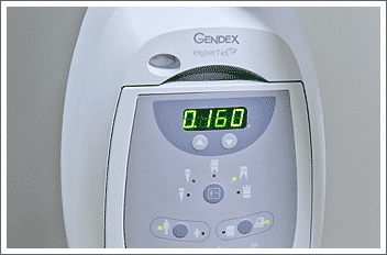
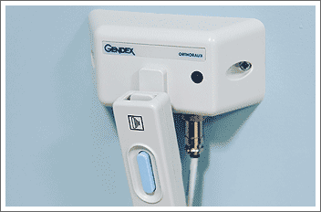
GENTEX EXPERT intra-oral x-ray unit
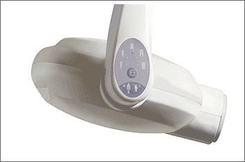
Expert DC with GXS-700 software
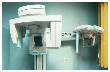
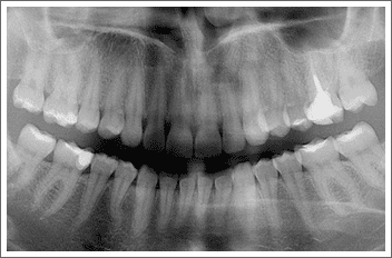
GENDEX (Orthoralix 9200DDE)
A single panoramic picture is sufficient to show the whole dentition and plan broadly understood dental treatment. It is worth noticing that in highly-developed countries, such regular tests are obligatory as they allow for elimination of any problems at their early stage before pain occurs, which often is indicative of a severe condition requiring costly treatment.
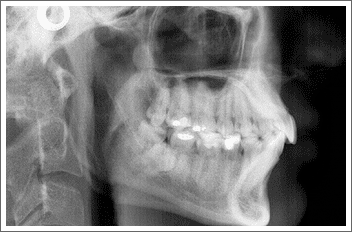
Cephalometric imaging module has a wide variety of applications, not only in orthognatic surgery. It is also used in orthodontics. MEDICO offers:
- panoramic x-rays for children (sensor movement is adapted to child’s anatomy, the radiation dose is lower)
- orthogonal dentition (without TM joint, perfect geometry and sharpness of the image of the dentition alone)
- semi-panoramic view (better geometry of the image, no spine shadow, radiation dose reduced by 50%)
- cephalometric (lateral) image of temporomandibular joints in occlusion and abduction
- temporomandibular joints in sagittal projection (DMF option)
- maxilliary sinus in sagittal projection (DMF option)
- maxilliary sinus in lateral projection (DMF option)
- orthogonal half dentition (DMF option)
- front teeth
2D imaging
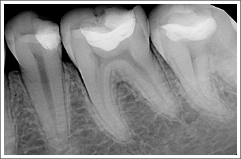
2D image, characteristic for pantomogram, intra-oral and occlusion pictures, is often “unreadable” due to “ghost shadows” - artefacts caused by bending of the x-ray film.
3D imaging
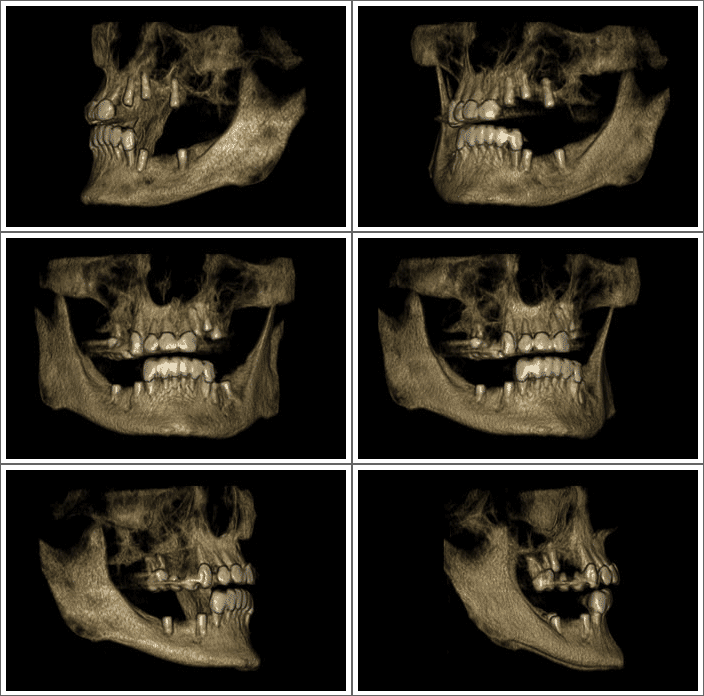
3D image shows the exact location of the nerves and sinuses, which - in the case of two-dimensional technologies - is never determined with a 100% confidence, thus posing the risk of damaging the nerve and causing paraesthesia.
Previously, only computer tomography could be used to obtain 3D picture of the facial skeleton. However, using an ordinary CAT scanner involved exposure to a high dose of harmful radiation.
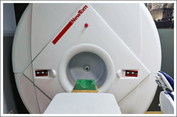
MEDICO's clinic is equipped with state-of-the-art Newtom cone beam CT scanner. Not only does it emit a small dose of radiation without burdening the patient's health, but also offers extraordinary precision. Out CAT scanner offers the highest, compared to ordinary tomography, resolution at considerably reduced dose of radiation.
Cone Beam Computed Tomography(CBCT) differs from ordinary tomography used in hospitals in terms of construction and algorithm generating the image.
Differences between Newtom and ordinary CAT scanner
Ordinary CAT scanner
The operation of an ordinary CAT scanner consists in computer calculation of the scanned area. This is done by means of detectors travelling along with an x-ray lamp around the scanned object. The number of projections necessary to obtain the cross-section is large, so is the number of rounds during which the radiation is emitted (from several dozens to several hundred).
CBCT scanner
In CBCT scanner the sensor is much larger and needs only one round to test the area of the size of the image generated. Therefore, the radiation dose to which the patient is exposed is incomparably smaller, which is a very important issue.
Parts of body that can be scanned
As suggested by the name, the radiation beam is cone-shaped, which enables radiation of the whole tested part and not only the surface, during a single round around the part concerned. Listed below are only some examples of the areas subject to scanning in dentistry:
- teeth
- sinuses
- temporomandibular joint
- sella
- facial soft tissue for the purpose of cephalometric measuring
Newtom CBCT device also allows for scanning other parts of the body, particularly important in orthopaedics, such as:
- 5 vertebral bodies of cervical vertebrae for the purpose of estimating bone age
- cervical spine
- feet
- wrists
Three-dimensional image
Images obtained during tomographic imaging in MEDICO is of the top quality; the process is very fast and the effects guarantee that the doctors will get a perfect picture of the patient’s anatomy, which is impossible in the case of 2D imaging which has always involved the risk or erroneous location of fibres, vessels or berves. Newtom allows for obtaining a 3D picture of any area of the facial skeleton and for obtaining 2D pantomograms. The data are stored in digital form and on a CD with a photo browser installed. Generated 3D model may be controlled manually. Dedicated software enables detailed observation of the current status and selection of the best therapy.
- elastography
- microcalcification
- USG of small organs: breasts, thyroid, testicles, female reproductive organs, osteoarticular and muscular system, chest and abdominal cavity.
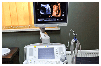
USG Toshiba Aplio M X
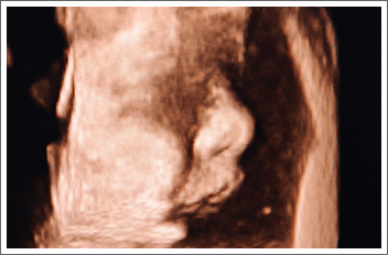
It is the newest ultrasound system of the next generation, ensuring top quality imaging. It consists of 3D and 4D modules, Doppler Duplex module (colour imaging of blood flows through blood vessels), and elastography module used to determine tissue flexibility and classify tissues based on the elasticity parameters (since 2010, this method has been used for the purpose of differentiating tumours; it may also help to determine the competence of certain organs, for instance of the uterine cervix during pregnancy).
Another imaging module featured by our Toshiba device is MicroPure allowing for detection of microcalcifications in breasts and small organs, which also - since 2010 - has been a breakthrough method in detecting and differentiating tumours. Both, elastography and microcalcification are innovative methods that MEDICO proudly boasts to have.


